Accurate positioning of the components is an important factor in the stability and longevity of a hip replacement. Modern pre-operative planning has evolved from basic 2D templating to detailed 3D virtual surgery. Combined with custom jigs and intra-operative imaging — part of the patient-specific technology I use for every joint replacement — the implant can be planned and positioned to within about a millimetre of the pre-operative plan.
How Pre-Operative Planning Has Advanced
Early hip replacement planning relied on 2D X-ray templating. While functional, this method had inherent limitations. The exact magnification of X-rays could vary, and bone rotation was difficult to account for. Surgeons often discovered during surgery that the pre-operative plan didn't match the patient's actual anatomy.
3D planning changed everything. Using CT or advanced imaging, I can now perform virtual hip replacement surgery weeks before you arrive at the hospital. I know the exact size, shape, position, and orientation of your individual hip anatomy. I can position the implant components in three dimensions and see how they interact before making a single cut.
"3D planning allows me to perform your surgery virtually before ever entering the operating theatre. Precision at this level was impossible just a decade ago."
- Dr Chien-Wen LiewWhat Happens During Pre-Operative Imaging
The 3D imaging process is remarkably simple and fast. You lie in the scanner for approximately 30 seconds while a special 3D scan is acquired. This is not an MRI requiring lengthy scanning times - it is a quick, high-resolution 3D image of your hip anatomy. The radiation exposure is minimal and comparable to a few standard X-rays.
Once the scan is complete, I perform comprehensive pre-operative planning. Using specialised software, I plan the exact size, shape, and position of each implant component specific to your anatomy. This virtual surgery is performed in three dimensions, allowing precise positioning that accounts for the patient's individual bone geometry, alignment, and hip anatomy.
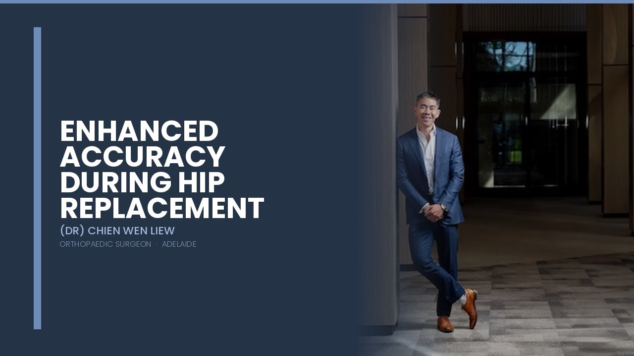
Dr Chien-Wen Liew explains the 3D planning and imaging checks he uses during hip replacement to compare implant position with the pre-operative plan.
Read transcript
0:00 · Introduction
We're going to go over what imaging modalities that we perform during hip replacement surgery. Now, if you're one of my patients or you've read a little bit through our website, you'll know that.
0:21 · Direct Anterior Approach
I use something called the direct anterior approach, and that is the preferred method for me of how I approach the hip. And that is only the approach and that encompasses how I actually access the hit. And we do that without cutting or detaching any of the muscles or tendons.
0:37 · Why we use advanced imaging
Now, the imaging modalities that we do surrounding the hip replacement to me are of critical importance. It gives us confidence in where we place the implant, but also allows us to pre-plan where the hip replacement should be. To match the implant to your anatomy as closely as possible now,
0:57 · Before the surgery
before the surgery, we would have gone through a few things together and we would have reviewed your pre-operative x-rays together. Now, what we do from there is, once we have booked the surgery, we organise a three-dimensional scan. Now, the three-dimensional scan that we perform is to look at the full morphology or shape of your hip and the surrounding structures. It gives us all of the dimensions of the acetabulum, which is the socket, the femoral head, which is the ball and the shape. The twist and the changes in the anatomy of the proximal femur, or the top of your hip bone.
1:37 · Why we plan in advance for THR surgery
Now, ultimately, what that means is that we do a lot of planning of the surgery before the actual surgery date. And it allows me to essentially perform the surgery on our computer many weeks before you actually come in for your surgery. It gives me the confidence to know that all of the anatomical aspects of the hip are known before we enter. And if we have any sort of abnormalities there.
We have a definite plan on what we need to do to aim for the best result for you. Now, this three-dimensional scan and the plan that I create is something that we then use to create a master plan for your hip.
2:13 · During surgery - technology
Where in Switzerland, a special jig is made, where we place that onto the hip during surgery. So that it allows us to plan for everything very accurately and execute it exactly as we planned now. Like anything, we never take anything for granted, and we're always measuring multiple times before we do anything. Now, during surgery, we do several things to ensure that we're extremely accurate with the position and size of the implant and how we place everything.
So it relates perfectly to how that hip is going to move, as every single person is built a little bit differently. When we go through the plans, we can see all of those differences and account for them at the time of surgery. And be knowledgeable about them so that there are no surprises on the day.
3:03 · Performing the surgery
Now, when we're performing the surgery, we place the acetabulum or the socket first, and then after that the femur. Once we've verified that the bony resections, which means the cuts on the bone, match exactly with our three-dimensional models, then we place all of the trial components inside the hip. Now, if you're one of my patients who have already been through surgery, you may have received something from me to show you what that looks like.
When we take that intraoperative image, I really prefer performing all of my operations with my patients. Supine, which means you're lying flat on your back comfortably during surgery.
3:43 · What kind of bed is used during surgery for THR
We use a very specialised bed during surgery, which is composed of carbon fibre and that allows me to image your hip during surgery. I feel that this is an integral part to the quality assurance that we have for positioning the hip. Prosthesis, as it allows us to see all of those elements that we may not be seeing during surgery, just by looking at the bone. And is a secondary check to how everything is positioned so that we can make sure that what we have placed compares perfectly to what we have planned.
4:15 · Optimising Accuracy during surgery
We do that imaging during the surgery so that we can make very small changes to the position of the implant with the final implantation of the device. So that things like leg length and position of the implant are compared against our pre-operative plan, once that's performed, we also do tests on range of movement, stability, as well as how all your soft tissues feel with the new hip replacement in there, so there are multiple checks on how we place that hip replacement before we close up the wound.
4:47 · Aims of surgery - THR
My aim is to ensure that once we have finished with the surgery, there is no need to go in and adjust any of the implants ever again. And that's really the aim of giving you a forgotten joint and one that will hopefully last an extremely long time.
I hope this has given you a little bit of insights into the imaging modalities that we use for a hip replacement. If you have any questions, please leave them in the comments below and I'll try and get back to you through that. Or else, contact our rooms for more information. In the meantime, please subscribe to our channel and I look forward to seeing you again for the next video.
Transcript edited lightly for readability.
Manufacturing Precision Tools for Your Hip
After virtual planning is complete, a custom jig - a precision surgical guide - is manufactured specifically for your anatomy. This jig is a perfect fit with your individual bone surfaces and femoral canal. It enables surgeons to prepare your hip bones with unprecedented accuracy, ensuring that the implant components are positioned exactly as planned.
Every patient gets a unique jig. This patient-specific technology is expensive and time-consuming to produce, but the accuracy it provides is invaluable. No two hips are identical, and no two jigs are identical. Each is custom-made to optimise accuracy for that specific patient's anatomy.
Real-Time Imaging During Surgery
Even with 3D planning and custom jigs, I verify implant positioning during surgery using intra-operative X-ray imaging. Our surgical table allows real-time X-ray scanning without repositioning the patient. This allows me to confirm that the implants are positioned exactly as planned before final fixation.
This intra-operative check is invaluable. If positioning is not optimal, I can adjust before the implants are permanently secured. This additional layer of quality control ensures that the final implant positioning meets my exacting standards for precision and alignment.
Why Every Patient Deserves This Approach
You might ask: why plan 3D for every patient? Don't some hips have "normal" anatomy that doesn't require special planning? The answer is that we cannot predict which hips will have anatomical variations. Approximately 40% of patients have some kind of anatomical abnormality or variant that requires additional pre-operative planning to ensure optimal positioning.
Because I cannot predict which patients will have these variations, I perform 3D planning and patient-specific technology for every hip replacement. This universal approach ensures that every patient receives the same exacting level of precision, regardless of whether their anatomy is straightforward or complex.

Orthopaedic Surgeon, Adelaide
Frequently Asked Questions
Download Planning Technology Guide
A detailed explanation of 3D planning, patient-specific jigs, and precision imaging in hip replacement.